
Introduction
A Stereo Microscope, also known as a Dissecting Microscope, is a low-magnification optical microscope designed for observing specimens in three dimensions. Unlike compound microscopes, it uses reflected light and two separate optical paths to provide depth perception, making it ideal for examining surface details of solid objects.
Principle
The stereo microscope operates on the principle of binocular vision. It uses two separate optical paths with two objectives and eyepieces, each angled slightly apart (typically 10–12°), allowing each eye to view the specimen from a different perspective. This creates a three-dimensional image through the phenomenon of stereopsis.
Working Procedure
Setup: Place the microscope on a stable surface and ensure proper lighting.
Specimen Placement: Place the sample on the stage using clips or plates.
Illumination: Use incident light for opaque specimens or transmitted light for translucent ones.
Magnification Adjustment: Adjust zoom or switch eyepieces to change magnification (typically 5x–250x).
Focus: Use coarse and fine focus knobs to sharpen the image.
Observation: View the specimen through both eyepieces for a 3D visualization.
Documentation: Capture images using an attached digital camera if available.
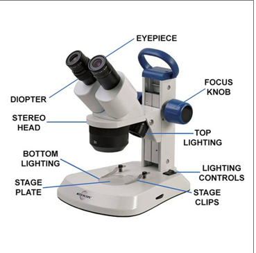
Picture of Stereo Microscope
Forensic Significance (Division-wise)
1. Biology Division
Examination of insects in forensic entomology.
Analysis of Hair, Fibers, and plant materials.
Identification of larvae or eggs in decomposition studies.
2. Physics Division
Inspection of tool marks, fracture surfaces, and glass fragments.
Analysis of gunshot residue (GSR) patterns and primer residues.
3. Questioned Documents Division
Detection of erasures, overwriting, and ink differences.
Examination of paper texture, indentations, and printing techniques.
4. Ballistics Division
Study of bullet striations, cartridge cases, and firing pin impressions.
Comparison of projectile surfaces for matching with firearms.
5. Entomology Division
Identification of insect species from partial remains.
Estimation of postmortem interval (PMI) using insect development stages.
Advanced Versions
Modern stereo microscopes come with:
Digital imaging systems for real-time 3D visualization.
LED illumination with adjustable brightness and colour temperature.
Motorized zoom and focus for precision control.
Ergonomic designs for prolonged use in forensic labs.
Software integration for image analysis and documentation.
Examples include:
BRESSER Advance ICD Zoom Stereo Microscope (10x–160x)
Radical RSMR Series with motorized focus and dark-field contrast
Conclusion
The stereo microscope is an indispensable tool in forensic science, offering unparalleled depth perception and surface detail analysis. Its versatility across divisions—from biology to ballistics—makes it vital for evidence examination, crime scene reconstruction, and legal investigations. With advancements in optics and digital integration, stereo microscopes continue to evolve, enhancing forensic capabilities and accuracy.
References
1. A review paper on stereomicroscope:
https://www.researchgate.net/publication/379383761_A_review_on_stereomicroscope
2. Wikipedia: https://en.wikipedia.org/wiki/Stereo_microscope